I’m excited to share the following publication from my colleagues at Neurovision Imaging & Cedars Sinai Medical Center. This paper represents a ton of work, from people all over the world. I’m also happy to say I had a chance to play a very small role in supporting the efforts!
This research continues to advance the work of attempting identification of Alzheimer’s via the retina. If we can continue to refine this approach, someday, people will be able to have frequent checkups to determine whether they are approaching a risk of Alzheimers – early enough for intervention to occur.
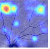
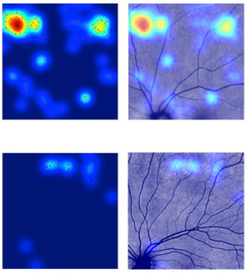
For the human trials, images were obtained of the retina, using a custom modified confocal ophthalmoscope. The retinal imager obtains images from the patient’s eye, and the images are then analyzed for a number of potential plaque sites (those sites which may contain AB plaques). If enough sites, and or site density is found, the patient indication shows higher, whereas a control would show less grouping and/or less sites. This is a simplified explanation of course, and a ton of work went into figuring out how to best identify these sites. Here’s an image of such a patient. Int his image the heat map indicates the grouping and intensity of the AB plaque locations.
Hats off to Maya & Yosef Koronyo, the team at Neurovision Imaging, and the folks at Cedars for this awesome work!
(Ref) https://insight.jci.org/articles/view/93621
-Austin