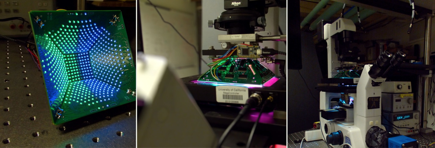
The Waller Lab recently had a demo announcement of their awesome implementation of computational imaging. They have some examples on their lytro page, which look really cool! This got me thinking about the last 10 years or so, and how the combination of image capture with control over input and output image fields, has grown into a new force in microscopy today.
It’s interesting to consider what techniques fit into the field of computational imaging. Optical microscopy has pushed into an ever smaller domain of resolution, to the point that Abbe’s laws need to be avoided in order to pull resolution out of an image. Consider the initial techniques used for this – phase, DIC, confocal, TIRF, and the like are contrast enhancement techniques of enhancing an image’s contrast, by either increasing the signal intensity, or by reducing the surrounding noise/background intensity. Beginning with the Apotome, and maybe earlier, with deconvolution, a combination of using multiple image samples with post-capture image processing, to gather more information than can be obtained by a single image, started to emerge.
Building on these first techniques, we can see further sampling methods in the optical domain, like STORM/PALM, where optical control over emission is coupled with high sampling rates, and back-end processing, to statistically avoid Abbe’s laws. I’d imagine FLIM falls into this category as well, using time and multiple images, again to extract more data than may be obtained by a single sample. Non-microscopy applications for yet another method in this domain, the employment of coded apertures, similar to this work from MIT, are growing in a number of imaging fields.
This combination, of computing power + controlled image fields, can be employed to do all sorts of as-yet untouched things with a conventional microscope. I can imagine that very soon, we will see a few of the following products available for use:
- phase contrast, and fluorescent, multi-capture resolution enhancement systems, all built into an “off the shelf” microscope
- angular changes to fluorescence excitation, used to improve resolution
- wavelength-based restriction inside an image field, to eliminate the potential for crosstalk in emission
- multiple excitation power sampling, to pull greater dynamic range from images
- multiple cameras used with restricting apertures, used to pull focus information, or multiple focal fields, from a single position in a sample
These are only a few ideas where this can go, but the general path I can see in the future is lower cost components, which are leveraged to do better and better work, which gives more imaging power to a greater number of researchers. At least – that’s a future I’d like to see 🙂
-Austin